70 Year-old Female with Unilateral Lower Extremity Swelling
Author: Ben Caviston, MD PGY-2
Peer Reviewers: Lee LaRavia, DO; Dan Kaminstein, MD
Learning Objectives:
Discuss the differential of unilateral lower extremity edema
Discuss use of POCUS in the workup
Discuss the US characteristics/findings associated with DVT
Review of literature related to POCUS for diagnosis of DVT
Case Presentation: H&P and Differential
70F hx of Endometrial Carcinoma presenting with Right lower extremity swelling for
one week now today with additional malaise, nausea and vomiting, No SOB or CP
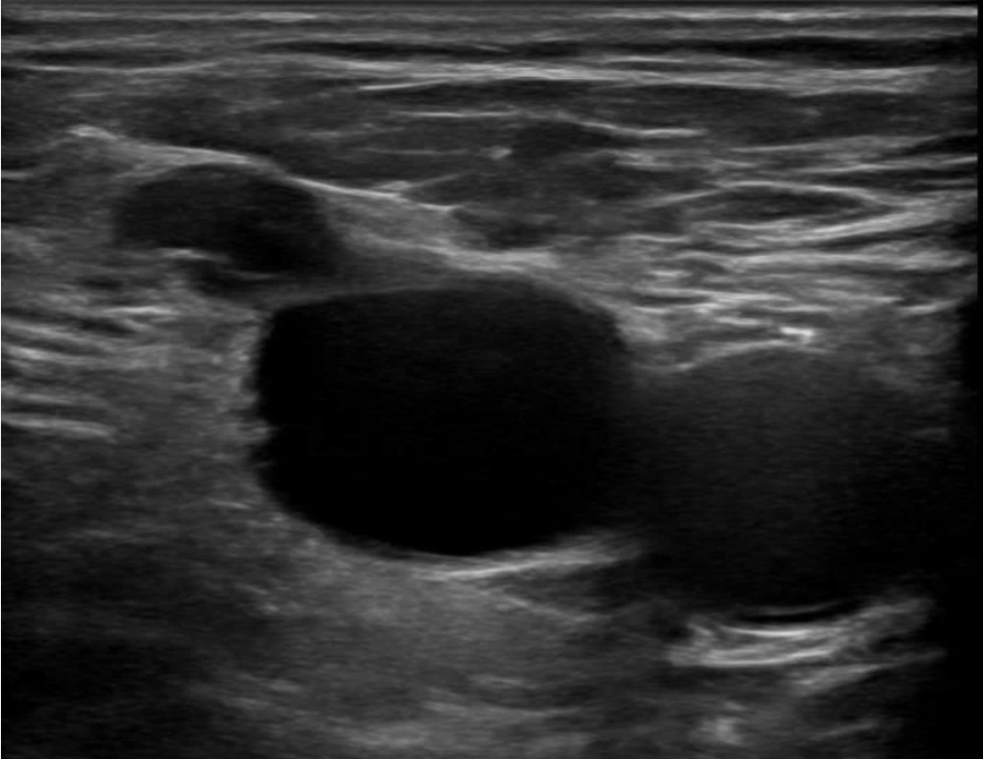
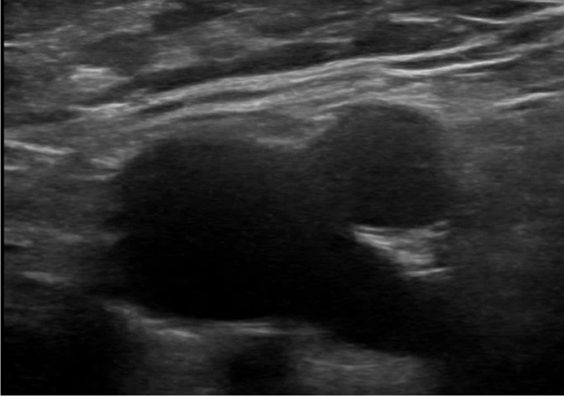
Begin scanning just below inguinal ligament in transverse axis, slide inferiorly
1.Identify the Femoral-Saphenous Junction
2.Identify the Femoral-Deep Femoral Junction
DVT POCUS Technique
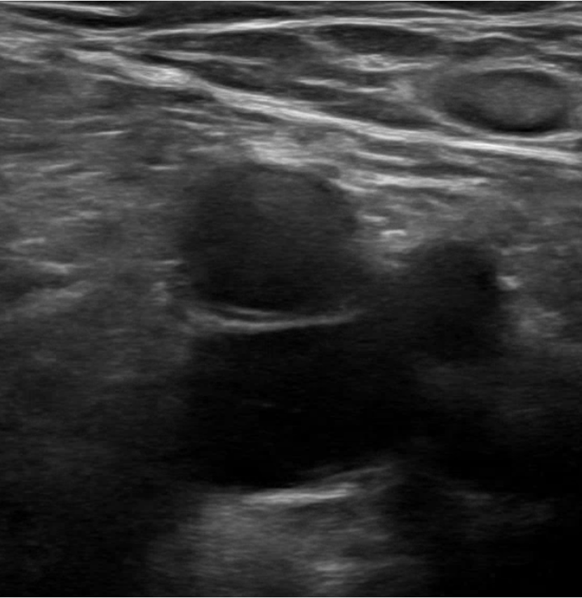
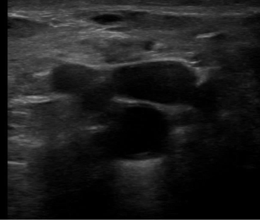
Zone-2 Popliteal Fossa
Knee partially flexed
Identify the Popliteal Vein
1.Identify the Popliteal Trifurcation
Splits into:
Peroneal Vein
Anterior Tibial Vein
Posterior Tibial Vein
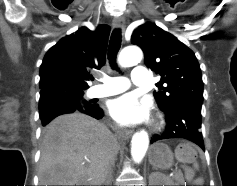
Diagnosis and Case Disposition
CTA showed Occlusive PE of the Right Superior Pulmonary Artery
Heparin drip started at 13:37 after POCUS
Radiology read of CTA 14:46
Recognizing DVT on POCUS allowed heparin to be initiated >1hr sooner
Literature Review
Bernardi et al 2008; 2465 patients randomized to either 2-Point Ultrasonography Plus
D-Dimer (Repeat US in 1wk if positive) vs Whole-Leg Doppler Ultrasonography
2 Point: Common femoral + Popliteal
Both strategies equivalent in DVT detection
Crisp et al 2015; ED POCUS 2 Point Compression vs Radiology Venous Duplex
ED physicians received 10min of training and used Portable Vascular Access Ultrasound
199 Patients and 45 w/DVTs all were identified by ED POCUS ( Sensitivity 100% )
Adhikari et al 2014; 6.3% of ED patients with suspected deep venous thrombosis had
isolated thrombi in proximal veins other than Common Femoral and Popliteal
Highlights importance of visualizing Femoral+Deep Femoral in addition to Common Femoral
Take Away Points
POCUS is highly sensitive and specific for DVT
Literature suggests similar Sensitivity as Radiology Whole Leg Venous Duplex
DVT most likely to form at branch points
Recommend Use of 2-Zone as opposed to 2-Point Compression
Ensuring visualization of the Deep Femoral Vein improves sensitivity
Consider routine use of POCUS in patients with signs of DVT or suspected PE
References
Adhikari S, Zeger W, Thom C, Fields JM. Isolated Deep Venous Thrombosis: Implications
for 2-Point Compression Ultrasonography of the Lower Extremity. Ann Emerg Med. 2015
Sep;66(3):262-6. doi: 10.1016/j.annemergmed.2014.10.032. Epub 2014 Nov 20. PMID: 25465473.
Bernardi E, et al; Erasmus Study Group. Serial 2-point ultrasonography plus D-dimer
vs whole-leg color-coded Doppler ultrasonography for diagnosing suspected symptomatic
deep vein thrombosis: a randomized controlled trial. JAMA. 2008 Oct 8;300(14):1653-9.
doi: 10.1001/jama.300.14.1653. PMID: 18840838.
Crisp JG, Lovato LM, Jang TB. Compression ultrasonography of the lower extremity with
portable vascular ultrasonography can accurately detect deep venous thrombosis in
the emergency department. Ann Emerg Med. 2010 Dec;56(6):601-10. doi: 10.1016/j.annemergmed.2010.07.010.
Epub 2010 Sep 22. PMID: 20864215.