January 2024
19 Year-old Female with Left Flank Pain, Nausea and Vomiting
Author: Ben Caviston, MD PGY-2
Peer Reviewers: Lee LaRavia, DO; Dan Kaminstein, MD; W. David Wynn, MD
Learning Objectives:
- List/discuss DDX of Left Flank Pain
- Discuss use of US in the workup
- Characterize US findings associated Nephrolithiasis
- Review of literature related to ED Renal POCUS
- Recognize the dangers of Anchoring bias and Bounce-backs
Case Presentation: H&P and Differential
- 19F presents with Left flank pain, dysuria, N/V. Seen 5d prior for similar, diagnosed with UTI prescribed Nitrofurantoin, symptoms worsening since
- T: 37.9 °C (Oral) HR: 149(Peripheral) RR: 30 BP: 120/73 SpO2: 99%
- Exam: Uncomfortable Appearing, LUQ/LLQ Tenderness, +Left CVA Tenderness
- DDx: Pyelonephritis, UTI, Urosepsis, GC/chlamydia, TOA, Pregnancy, Ectopic, Nephrolithiasis, Septic Stone, Diverticulitis, Gastritis/PUD, MSK, Thoracic Pathology, AAA, Splenic Rupture
Case Presentation cont: Initial Workup
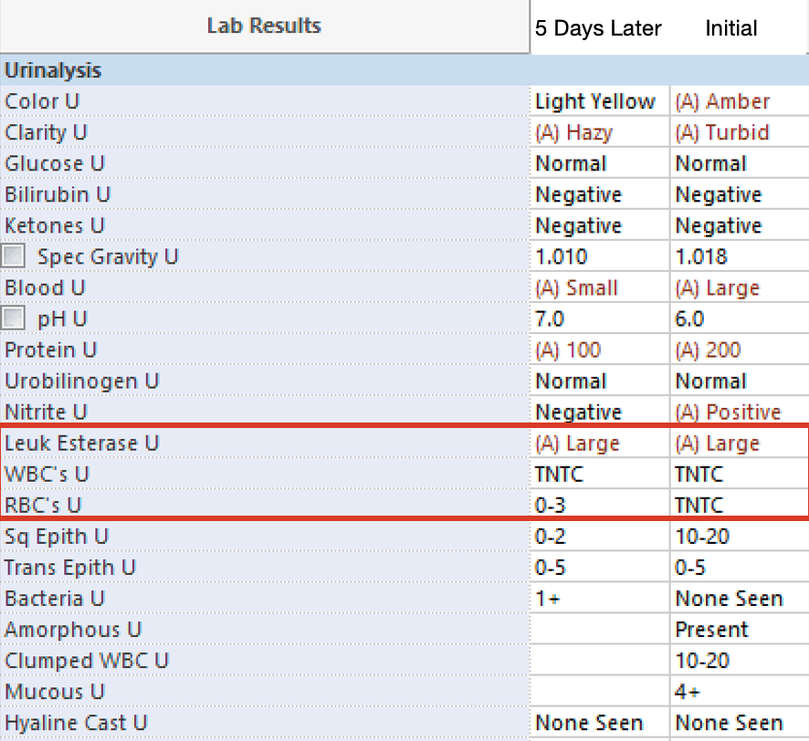
- CBC, CMP, BCx, UA, Hcg, BCx
- Urine Pregnancy Negative
- WBC 11.6; 79% segs
- CRP 34
- Cr 0.75 → 1.24
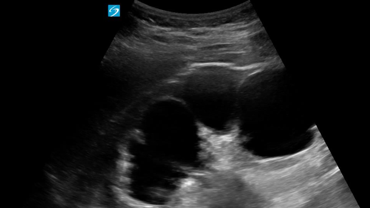
POCUS Images
POCUS QA
- Rib shadows can be overcome by angling probe between rib spaces
- Depth should be optimized
- Attempt visualization of proximal ureter
- Labels are always very helpful
Diagnosis and Disposition
Septic Stone
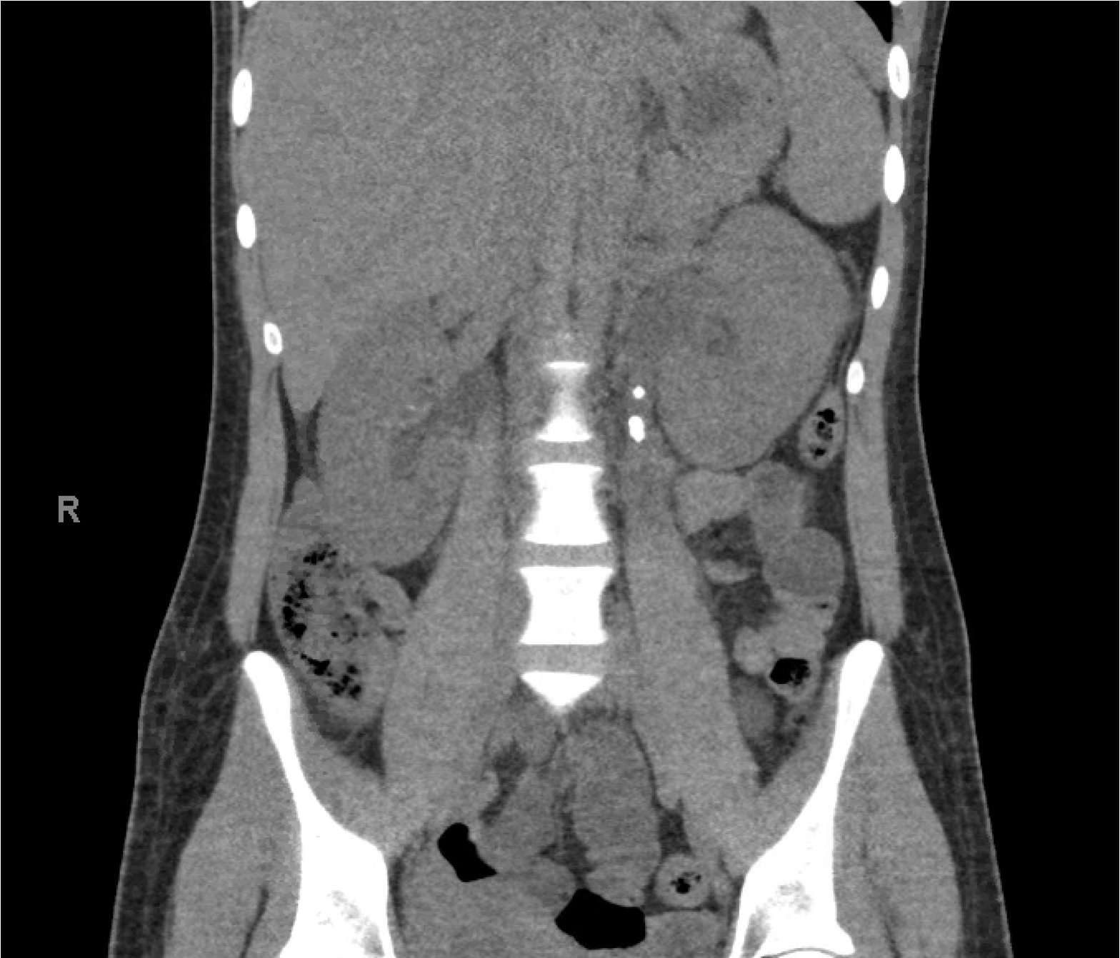
- Based on POCUS findings CT Stone Hunt Obtained
- Left hydroureteronephrosis
- Two obstructing Stones in Proximal Ureter
- Measuring 8 and 4mm
- Urology Consulted
- Lvl 2 to OR for Ureteral Stent Placement
Hydronephrosis Grading
MILD - Dilation of Renal Pelvis
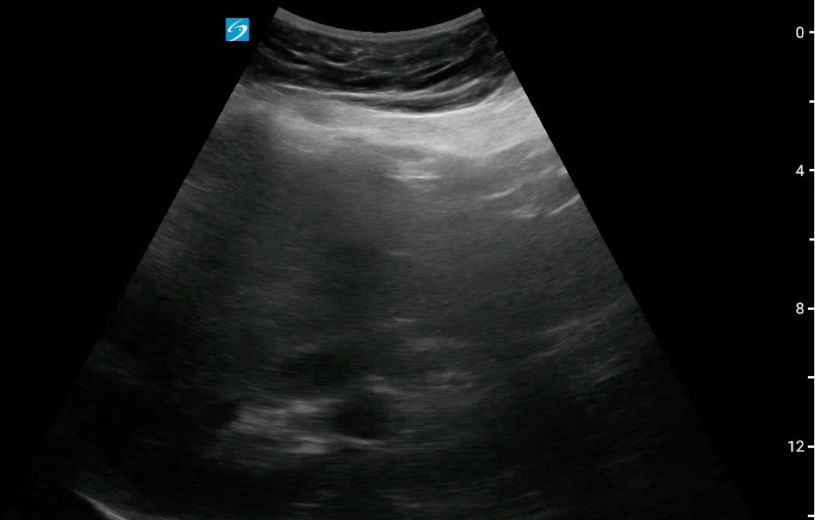
MODERATE - Dilation of Pelvis+Major Calyces
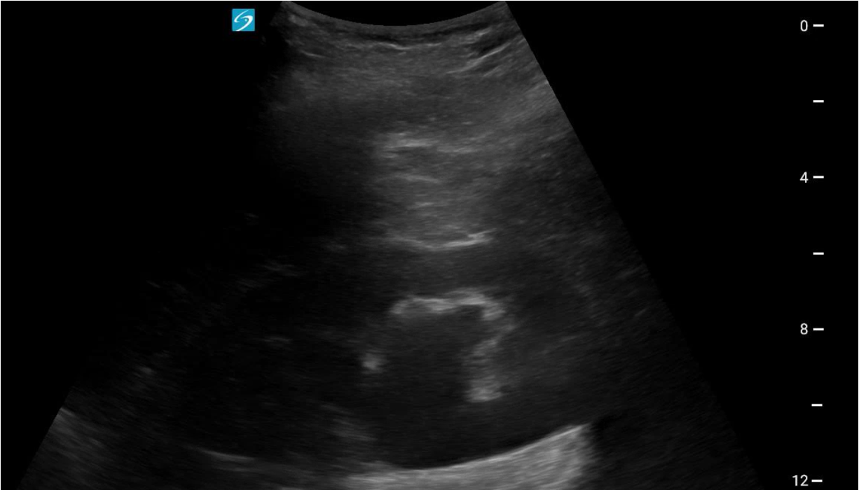
SEVERE - Dilation of Pelvis+Major Calyces +/- Cortical Thinning based on Chronicity
- Recommend use of simplified grading system Mild-Moderate-Severe
- No single consensus system exists, numerous exist
- Most recognized is SFU grade (I – IV) which was developed in prenatal US and unnecessarily complex for POCUS
Literature Review
- Smith-Bindman R et al (2014): POCUS vs. Rads US vs. CT in 2759 patients
- No difference between POCUS vs RUS; however less sensitive than CT
- ED stay 1.3hr Shorter, no significant difference in 30d complications
- Wong et al (2018) Systematic review of POCUS for renal colic
- POCUS had modest sensitivity of 0.70 (95% CI 0.67-0.73), specificity of 0.75 (95% CI 0.73-0.78) for any degree of hydronephrosis, performance better in setting of moderate severity or higher
- Taylor M et al. (2016) Ultrasonography for the prediction of urological surgical intervention
- Presence of a stone or moderate-severe Hydro Sensitivity 0.97 (CI 0.89-1) for surgical intervention specificity only 0.28
- Presence of both Stone and Hydro increased Specificity to 0.91 (CI 0.88-0.94) with +LR 2.94
- Sibley et al (2020) with binary interpretation Hydro vs no Hydro trainees with 25 scans had no difference compared with more experienced providers
Take Away Points
1. While POCUS has moderate sensitivity/specificity for Proximal Stones, has excellent sensitivity for need of Urologic Intervention
2. Applied in the correct patient may reduce Radiation and length of ER stay
3. Detection of moderate-severe hydro associated with need for intervention
4. Consider routine use of POCUS as a Screening Tool in low-risk patients
5. Maintain broad differential in ED bounce-backs and avoid anchoring
References
Sibley S, Roth N, Scott C, Rang L, White H, Sivilotti MLA, Bruder E. Point-of-care
ultrasound for the detection of hydronephrosis in emergency department patients with
suspected renal colic. Ultrasound J. 2020 Jun 8;12(1):31. Smith-Bindman R et
al.: Ultrasonography versus computed tomography for suspected nephrolithiasis. N Engl
J Med. 2014;371(12):1100-10.
Taylor M, Woo MY, Pageau P, McInnes MD, Watterson J, Thompson J, Perry JJ. Ultrasonography
for the prediction of urological surgical intervention in patients with renal colic.
Emerg Med J. 2016 Feb;33(2):118-23. doi: 10.1136/emermed-2014-204524.
Wong C et al. The accuracy and prognostic value of point-of-care ultrasound for nephrolithiasis
in the emergency department: a systematic review and meta-analysis. Acad Emerg Med.
2018;25(6):684-98