March 2026
20 Year-old Recent STI Treatment and Medically Induced Abortion Presenting to the ED for Vaginal Bleeding
Author: Man Preet Luthra, MD PGY-3
Peer Reviewers: Lee LaRavia, DO
Learning Objectives:
- List/discuss DDX for vaginal bleeding
- Discuss use of US in the workup of 1st trimester bleeding
- Discuss the US characteristics/findings associated with Molar Pregnancy
- Research specificity/sensitivities of detecting molar pregnancy
Case Presentation
- This is a 20-year-old female with a past medical history of chlamydia status post treatment of both herself and partner, recent diagnosed pregnancy in the 3rd week of January, medically induced abortion within the 1st week of February, that presents to the ED for worsening vaginal bleeding that has been ongoing since induction of medical abortion in the 1st week of February. (Today is April 7)
- Vitals: HR-112, BP-126/88, RR-16, T-98.6F, SpO2-100%
- Suprapubic tenderness, severe conjunctival pallor, light-headedness upon standing
- Urine Preg +
- DDx: Retained products of conception, Septic abortion, Ectopic Pregnancy, gestational trophoblastic disease (umbrella term)
POCUS Videos
Longitudinal Transabdominal : Cystic mass with no visible anatomical structures that represent a fetus (e.g heart or other organs).
Note: Forgot to put on doppler but would recommend.
Transverse Transabdominal: Cystic mass with no visible anatomical structures that represent a fetus (e.g heart). There is a hypoechoic pocket near the cervical canal. Note: Would adjust depth
Other Important Labs
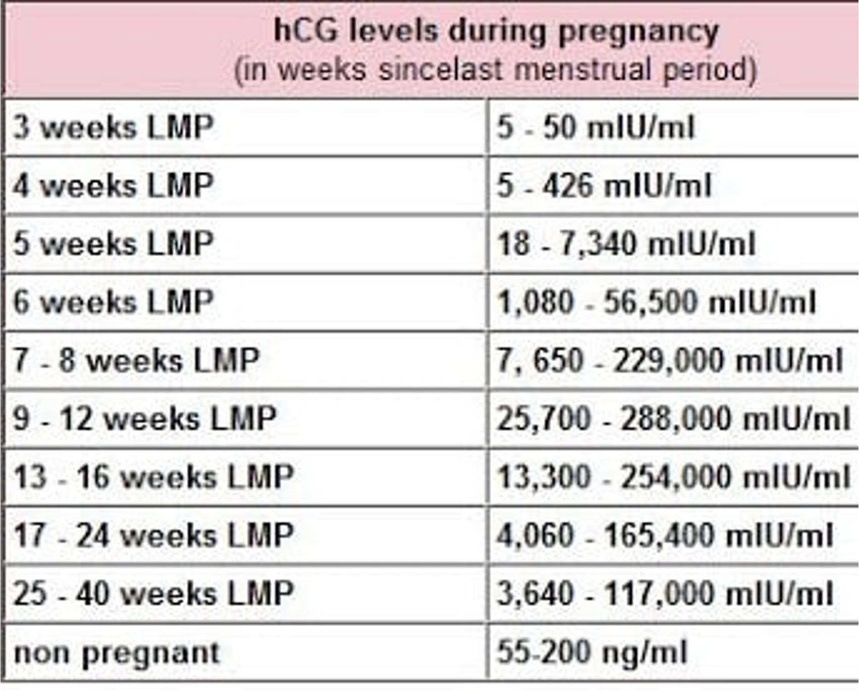
Hgb 6.9 (baseline 12.5), BHCG 266,000 at no more than 8 weeks pregnant by last LMP (End of 1st week of February).
HCG important for several reasons,
1.Discriminatory zone (level at which IUP should be visible with US)
- 6,000-6,500 for transabdominal US
- 1,000-2,000 for transvaginal US
2.Gestational age estimation
3.Assess for HCG in molar pregnancy
Diagnosis and Case Disposition
- 1. Transfusion of 2 units of PRBC s/p type and cross.
- 2. Confirmatory radiology performed transabdominal US was ordered.
- 3. Consult to OBGYN with Level 2 OR activation for D&C and admit by OBGYN.
- 4. Pathology report:
- 5. Patient had recurrence which indicated development of choriocarcinoma
Ultrasound Findings: CHM
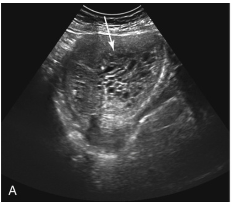
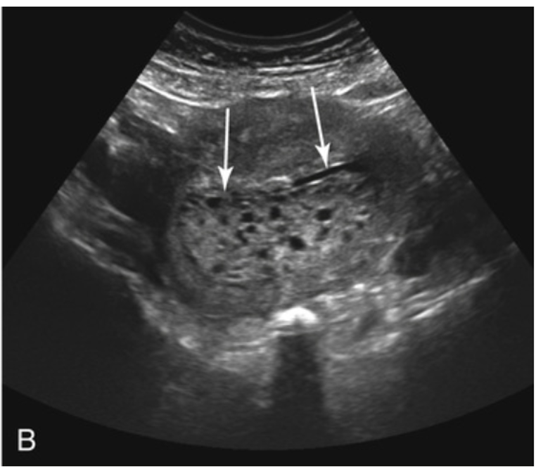
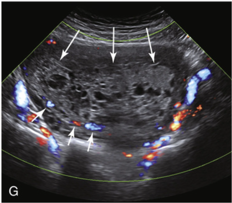
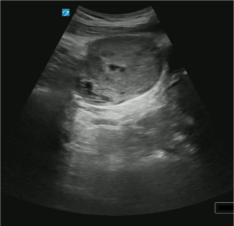
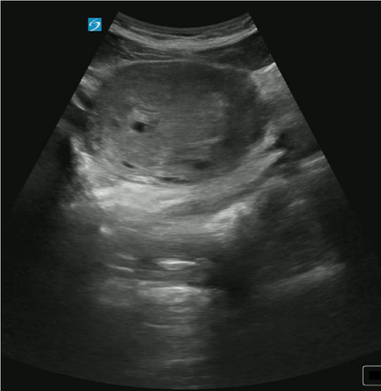
Ultrasound Findings for complete mole:
1.Large uterus with hyperechoic or isoechoic tissue
2.Multiple cysts of varying sizes but if villi are too small it may appear as a echogenic mass that appears solid
3.Doppler over the noninvasive mole with little to no blood flow in the molar tissue and flow in the periphery.
4.No fetal parts will be seen.
Ultrasound Findings: CHM
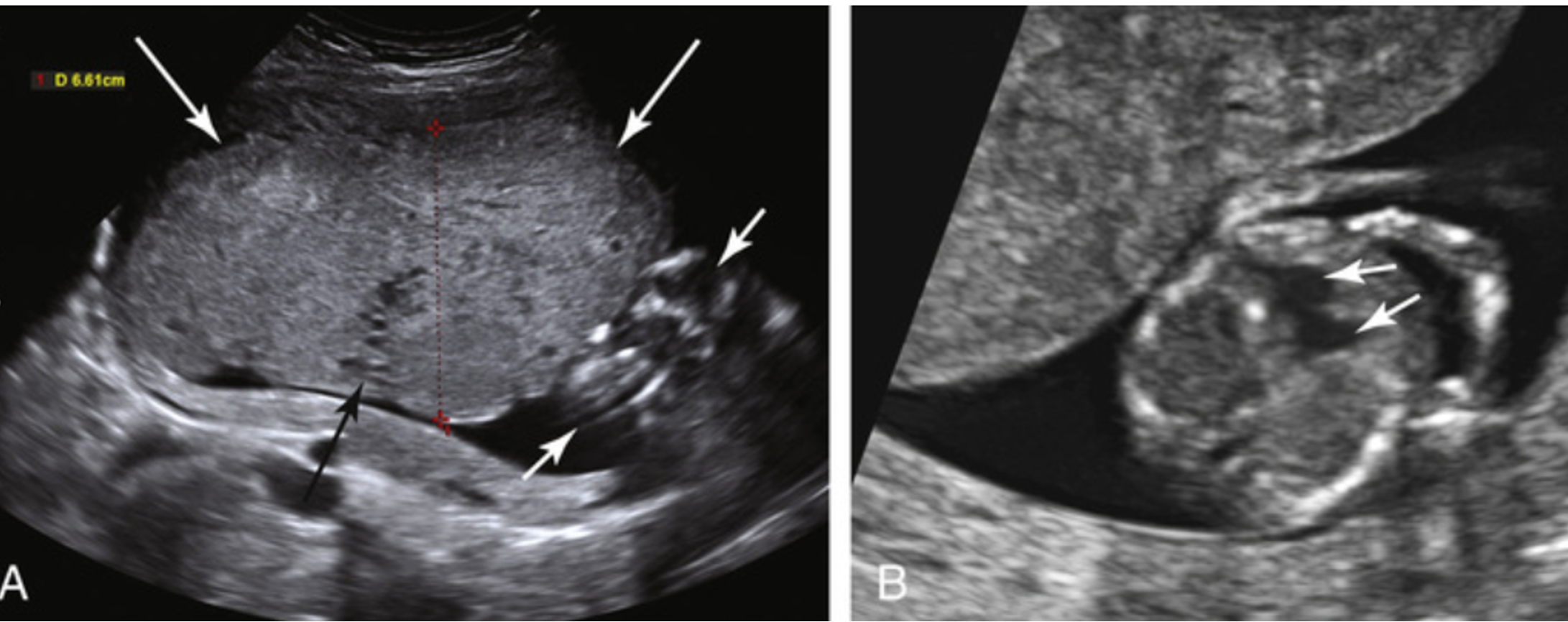
Ultrasound Findings PHM:
1.Large placenta with cystic changes.
2.Gestation sac is often present.
3.Variable identification of a fetus, but often times fetus has triploid karyotype which may or may not be due to the many anatomical anomalies.
Literature Review
- 1 in 700 pregnancies in the United States are complicated by a partial hydatidiform mole, whereas complete hydatidiform moles occur in 1 in 1500 pregnancies. 20% of complete moles develop into trophoblastic disease (e.g. choriocarcinoma) vs 5% of partial moles develop trophoblastic disease.3
- Complete mole form in an egg with no genetic material however form a diploid when two paternal spermatozoa fertilized the egg. Whereas partial mole is formed from an abnormal combination of an ovum and one or more spermatozoa. (>Diploid).
Literature Review
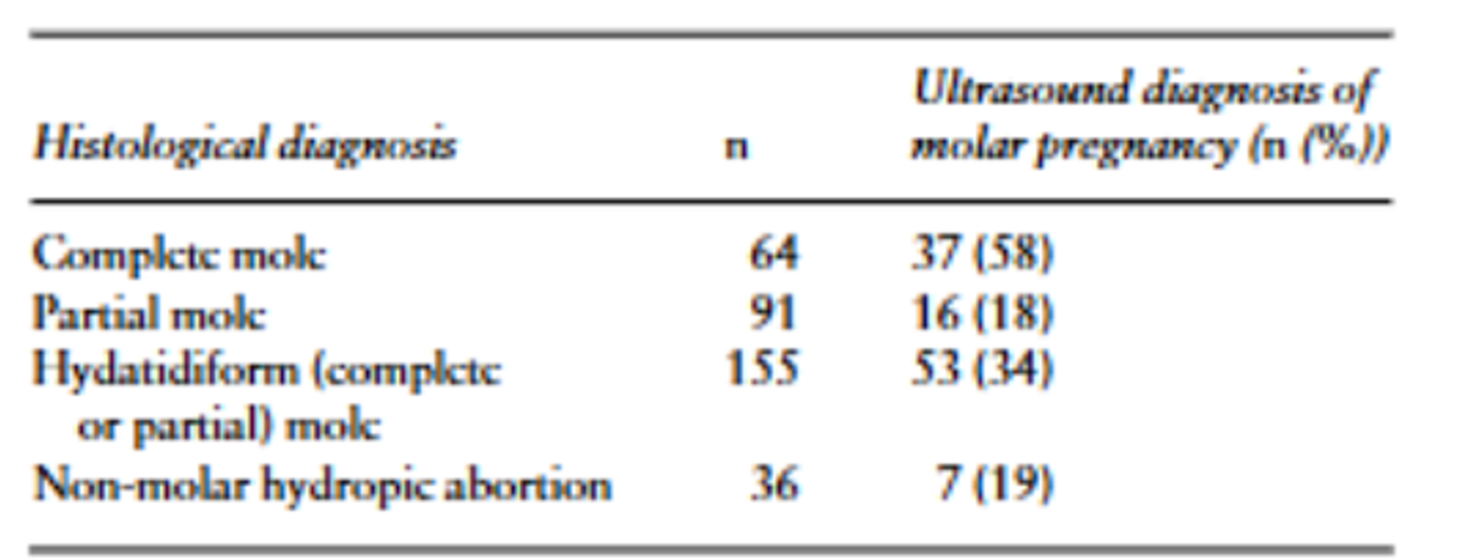
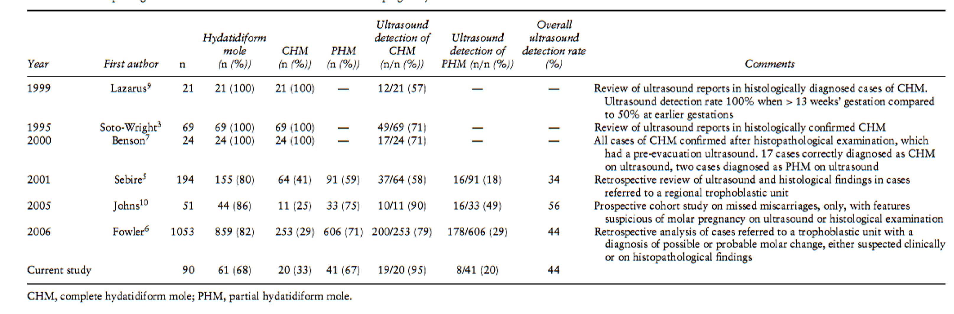
- Retrospective review of a 194 women that were referred to National Trophoblastic Disease Surveillance Centre over a 6-month period. (Sebire 2001)
Literature Review
- The sensitivity and positive predictive value for the ultrasound diagnosis of hydatidiform mole was 44% and 48%, respectively. For PHMs the respective values were 20% and 22% and for CHMs they were 95% and 40%.3
- Other studies reflect similar patterns (Kirk et al., 2007)
Take Away Points
1.Implement US in any 1st trimester vaginal bleeding.
2.Use the entire clinical picture to increase your likelihood of diagnosing molar pregnancy.
3.CHM is easier to detect than PHM with ultrasound.
4.Ultrasound detection for CHM is >80% (in some studies) while PHM is much lower.
5.Be aware of the βhCG discriminatory zone. βhCG level often exaggeratedly elevated for gestational age in CHM but less so in PHM making it a potentially unreliable marker just as in ectopic pregnancy.
References
1.American Pregnancy Association. (2012, April 26). What Does a high hCG Level Mean? American Pregnancy Association. https://americanpregnancy.org/getting-pregnant/hcg-levels/
2.Hertzberg, B. S., & Middleton, W. D. (2016). Ultrasound : the requisites (Third, Vol. 1, pp. 322–352). Elsevier. https://bookshelf.health.elsevier.com/reader/books/9780323314343/epubcfi/6/52[%3Bvnd.vst.idref%3Did_chp0014]!/4/2/20/2[s0115]/4[p0565]/2/1:20[bla%2Csti]
3.Kirk, E., Papageorghiou, A. T., Condous, G., Bottomley, C., & Bourne, T. (2007). The accuracy of first trimester ultrasound in the diagnosis of hydatidiform mole. Ultrasound in Obstetrics and Gynecology, 29(1), 70–75. https://doi.org/10.1002/uog.3875
4.Savage, J. L., Maturen, K. E., Mowers, E. L., Pasque, K. B., Wasnik, A. P., Dalton, V. K., & Bell, J. D. (2016). Sonographic diagnosis of partial versus complete molar pregnancy: A reappraisal. Journal of Clinical Ultrasound, 45(2), 72–78. https://doi.org/10.1002/jcu.22410
5.Sebire, N. J., Rees, H., Paradinas, F., Seckl, M., & Newlands, E. (2001). The diagnostic implications of routine ultrasound examination in histologically confirmed early molar pregnancies. Ultrasound in Obstetrics and Gynecology, 18(6), 662–665. https://doi.org/10.1046/j.0960-7692.2001.00589.x